Neurocloud SISCOM

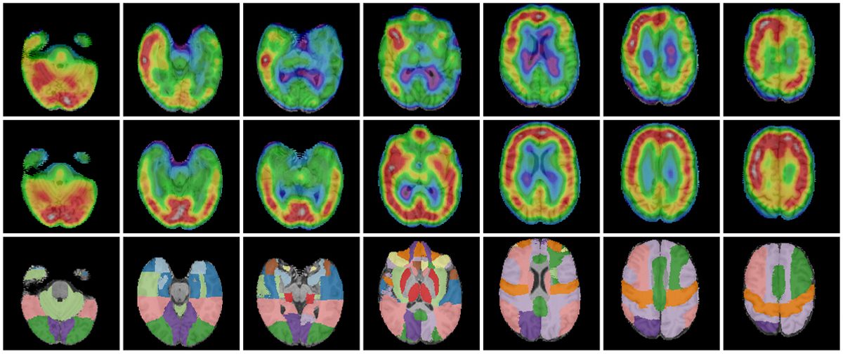
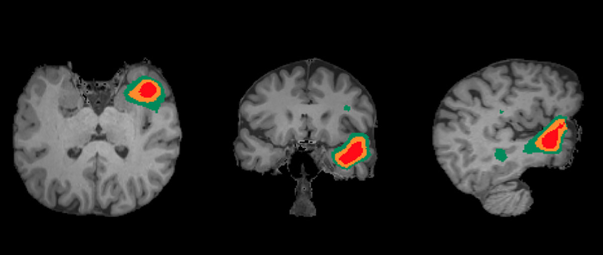
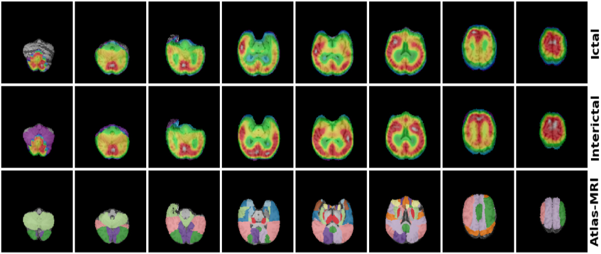
The software performs co-registration of SPECT images with the patient’s MRI. Subsequently, the subtraction of the interictal SPECT image is applied to the ictal image and the epileptogenic area is identified on the MRI image.
The SISCOM procedure is recommended by the EANM in the study of epilepsy, and provides a sensitivity greater than 80% and a specificity greater than 75% in the location of the epileptogenic focus.
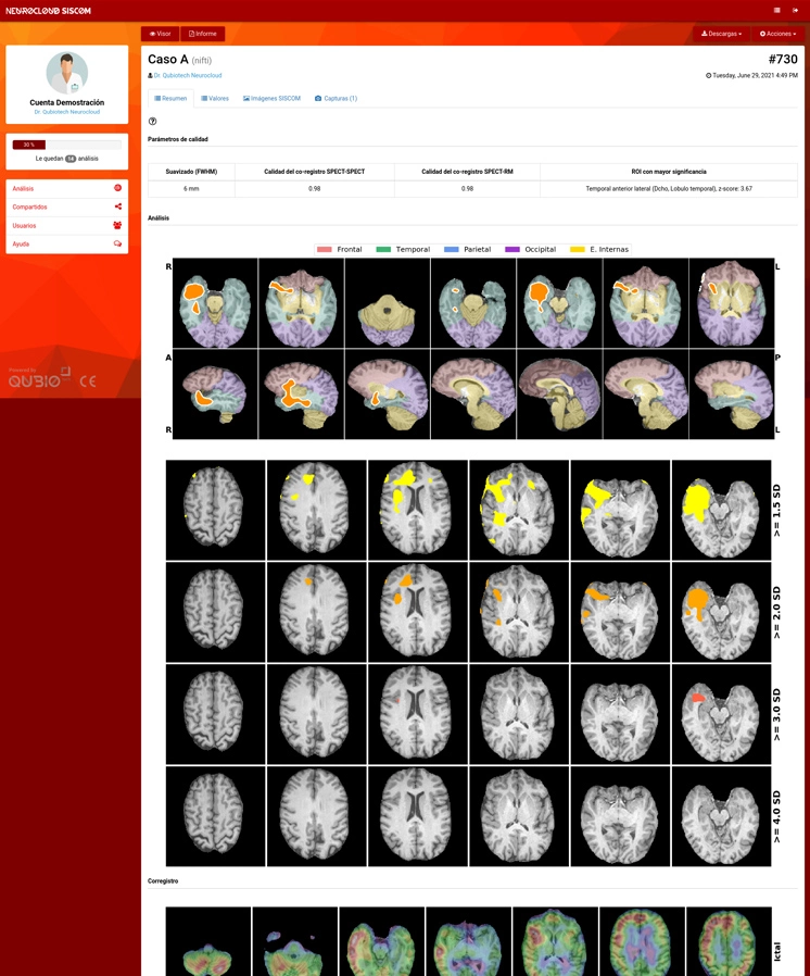
The results of Neurocloud – SISCOM consists of a parametric map of the voxels identified as an epileptogenic zone on MRI, data tables with the maximum Z-Score for each region of interest ( ROI) as well as the percentage of focus overlap and the SPECT-MRI fusion image.
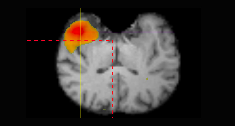
Help in epileptology
Identify the epileptogenic focus quickly and accurately.
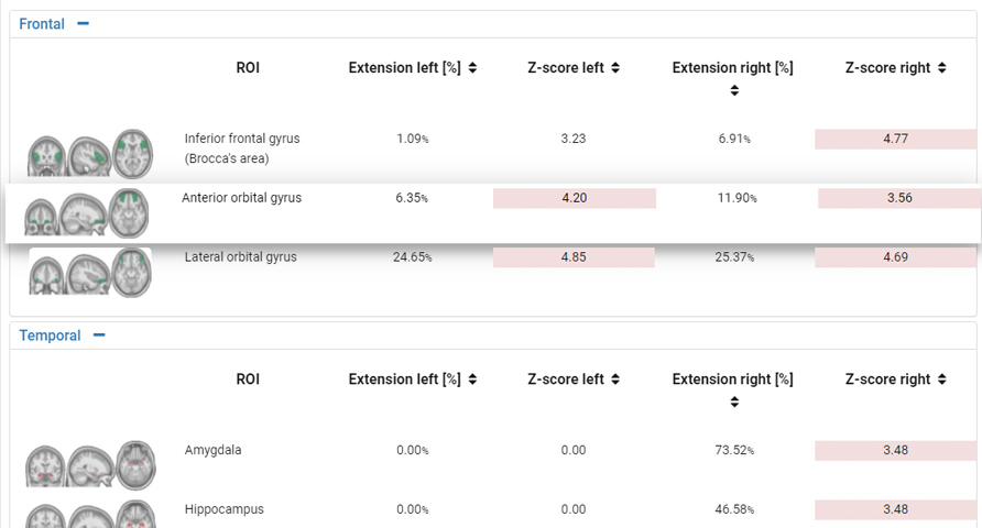
Help in neurology
The ictal SPECT subtraction procedure and MRI co-registration demonstrate a sensitivity greater than 80% and a specificity greater than 90% in the location of the epileptogenic focus.
Help in diagnosis and follow-up
Independent of the observer
Eliminates inter-observer differences between specialists. Enables you to move from a subjective working model to an objective model based on observer-independent data.
Automatic
Automatic processing and results in minutes. Connectivity with PACS systems for seamless integration with your workflow.
Sensitive
The SISCOM procedure provides a sensitivity above 80% in the location of the epileptogenic focus.
All in one
It integrates all the necessary resources for the diagnosis:
results in images, data tables and interactive viewer.
Neurocloud SISCOM: ICtal and interictal SPECT subtraction with MRI
> Co-registration of ictal and interictal SPECT> Subtraction. Ictal and interictal imaging> Visually identify the epileptogenic zone (EZ)
100% Compatible with your equipment and PACS
Fast, intuitive and fully automated
Invisible integration in clinical practice
Continuous and free updates
Continuous and personalized assistance
Hiring adapted to your needs
Clinically validated, CE marked
